


 大包装询价
大包装询价 产品介绍 评论(0)
宿主来源
Rabbit抗原名称
β-tubulin分子别名
TUBB; TUBB5; Tubulin beta-5 chain免疫原
Recombinant Protein细胞定位
IntracellularAccession
P07437克隆号
SDT-R014抗体类型
Rabbit mAb应用
ICFCM, IHC-P, ICC, WB反应种属 ?
Hu, Ms, Rt纯化方式
Protein A浓度
0.5mg/ml分子量
55kDa
标记
Unconjugated性状
Liquid缓冲体系
PBS, 40% Glycerol, 0.05%BSA, 0.03% Proclin 300
储存条件
12 months from date of receipt / reconstitution, 2 to 8 °C as supplied.
| 应用 | 稀释度 |
|---|---|
| ICFCM | 1:500 |
| WB | 1:1000-1:10000 |
| IHC-P | 1:1000 |
| ICC | 1:500 |
α- and β-tubulin polymerize into dynamic microtubules. In eukaryotes, microtubules are one of the major components of the cytoskeleton, and function in many processes, including structural support, intracellular transport, and DNA segregation. To form microtubules, the dimers of α- and β-tubulin bind to GTP and assemble onto the (+) ends of microtubules while in the GTP-bound state. The β-tubulin subunit is exposed on the plus end of the microtubule, while the α-tubulin subunit is exposed on the minus end. After the dimer is incorporated into the microtubule, the molecule of GTP bound to the β-tubulin subunit eventually hydrolyzes into GDP through inter-dimer contacts along the microtubule protofilament.
免疫印迹

WB result of β-Tubulin Rabbit mAb
Primary antibody: β-Tubulin Rabbit mAb at 1/1000 dilution
Lane 1: Hela whole cell lysate 20 µgSecondary antibody: Goat Anti-Rabbit IgG, (H+L), HRP conjugated at 1/10000 dilution
Predicted MW: 50 kDa
Observed MW: 52 kDa
econds
WB result of β-Tubulin Rabbit mAb
Primary antibody: β-Tubulin Rabbit mAb at 1/1000 dilution
Lane 1:Mouse brain lysate 20 µg
Lane 2: NIH/3T3 whole cell lysate 20 µgSecondary antibody: Goat Anti-Rabbit IgG, (H+L), HRP conjugated at 1/10000 dilution
Predicted MW: 50 kDa
Observed MW: 52 kDa
WB result of β-Tubulin Rabbit mAb
Primary antibody: β-Tubulin Rabbit mAb at 1/1000 dilution
Lane 1:rat brain lysate 20 µg
Secondary antibody: Goat Anti-Rabbit IgG, (H+L), HRP conjugated at 1/10000 dilution
Predicted MW: 50 kDa
Observed MW: 52 kDa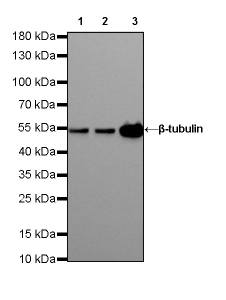
WB result of β-Tubulin Rabbit mAb
Primary antibody: β-Tubulin Rabbit mAb at 1/10000 dilution
Lane 1: HeLa whole cell lysate 20 µg
Lane 2: NIH/3T3 whole cell lysate 20 µg
Lane 3: rat brain lysate 20 µg
Secondary antibody: Goat Anti-Rabbit IgG, (H+L), HRP conjugated at 1/10000 dilution
Predicted MW: 50 kDa
Observed MW: 52 kDa
流式分析
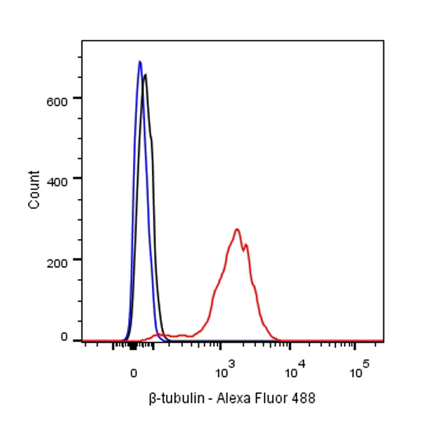
Flow cytometric analysis of HeLa cells labelling β-tubulin antibody at 1/500 (0.1ug) dilution/ (red) compared with a Rabbit monoclonal IgG (Black) isotype control and an unlabelled control (cells without incubation with primary antibody and secondary antibody) (Blue). Goat Anti-Rabbit IgG Alexa Fluor® 488 was used as the secondary antibody.
免疫组化

IHC shows positive staining in paraffin-embedded human testis.
Anti-β-tubulin antibody was used at 1/1000 dilution, followed by a Goat Anti-Rabbit IgG H&L (HRP) ready to use.
Counterstained with hematoxylin.
Heat mediated antigen retrieval with Tris/EDTA buffer pH9.0 was performed before commencing with IHC staining protocol.
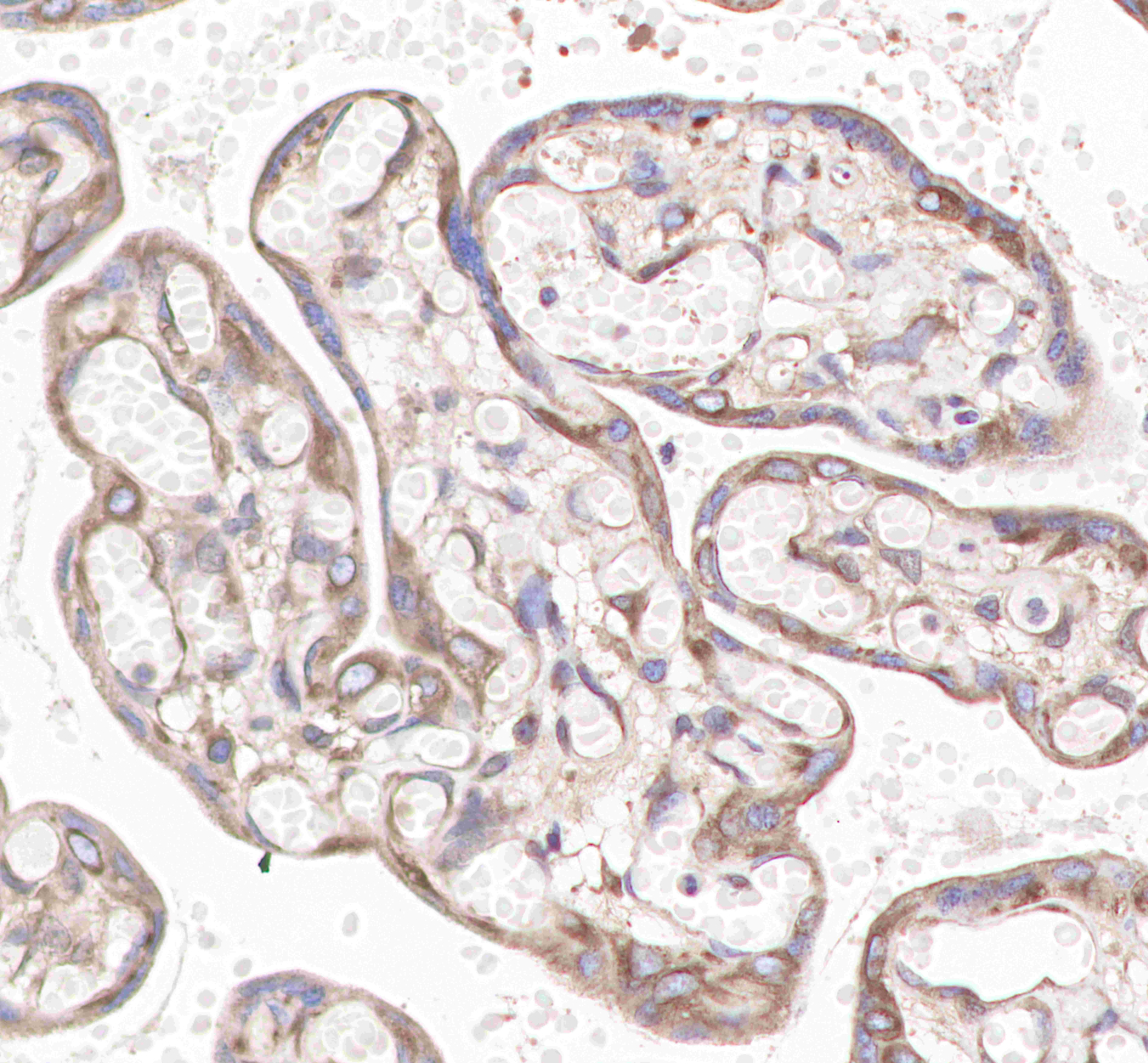
IHC shows positive staining in paraffin-embedded human placenta.
Anti-β-tubulin antibody was used at 1/1000 dilution, followed by a Goat Anti-Rabbit IgG H&L (HRP) ready to use.
Counterstained with hematoxylin.
Heat mediated antigen retrieval with Tris/EDTA buffer pH9.0 was performed before commencing with IHC staining protocol.

IHC shows positive staining in paraffin-embedded human cerebral cortex.
Anti-β-tubulin antibody was used at 1/1000 dilution, followed by a Goat Anti-Rabbit IgG H&L (HRP) ready to use.
Counterstained with hematoxylin.
Heat mediated antigen retrieval with Tris/EDTA buffer pH9.0 was performed before commencing with IHC staining protocol.
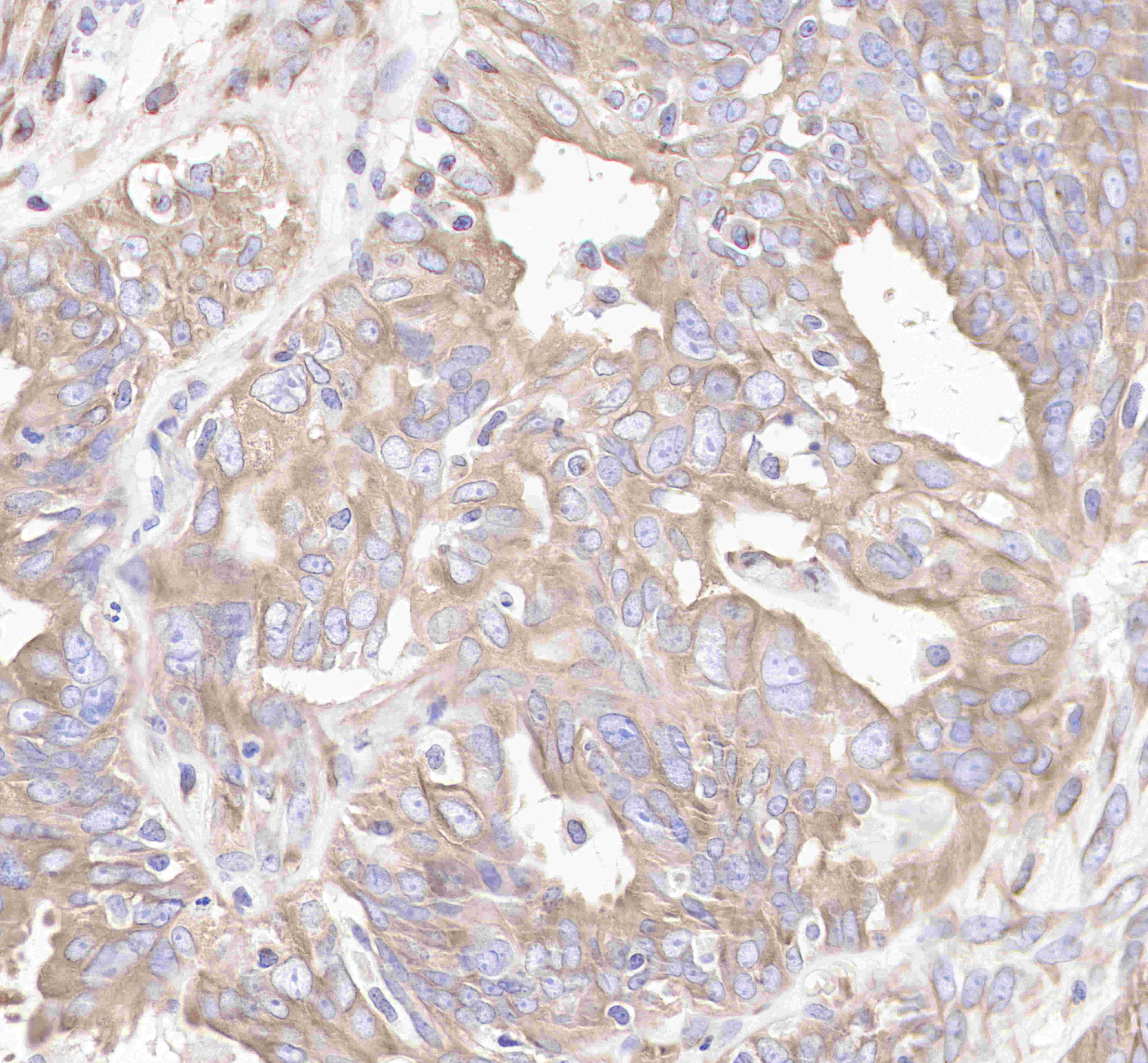
IHC shows positive staining in paraffin-embedded human colon cancer.
Anti-β-tubulin antibody was used at 1/1000 dilution, followed by a Goat Anti-Rabbit IgG H&L (HRP) ready to use.
Counterstained with hematoxylin.
Heat mediated antigen retrieval with Tris/EDTA buffer pH9.0 was performed before commencing with IHC staining protocol.
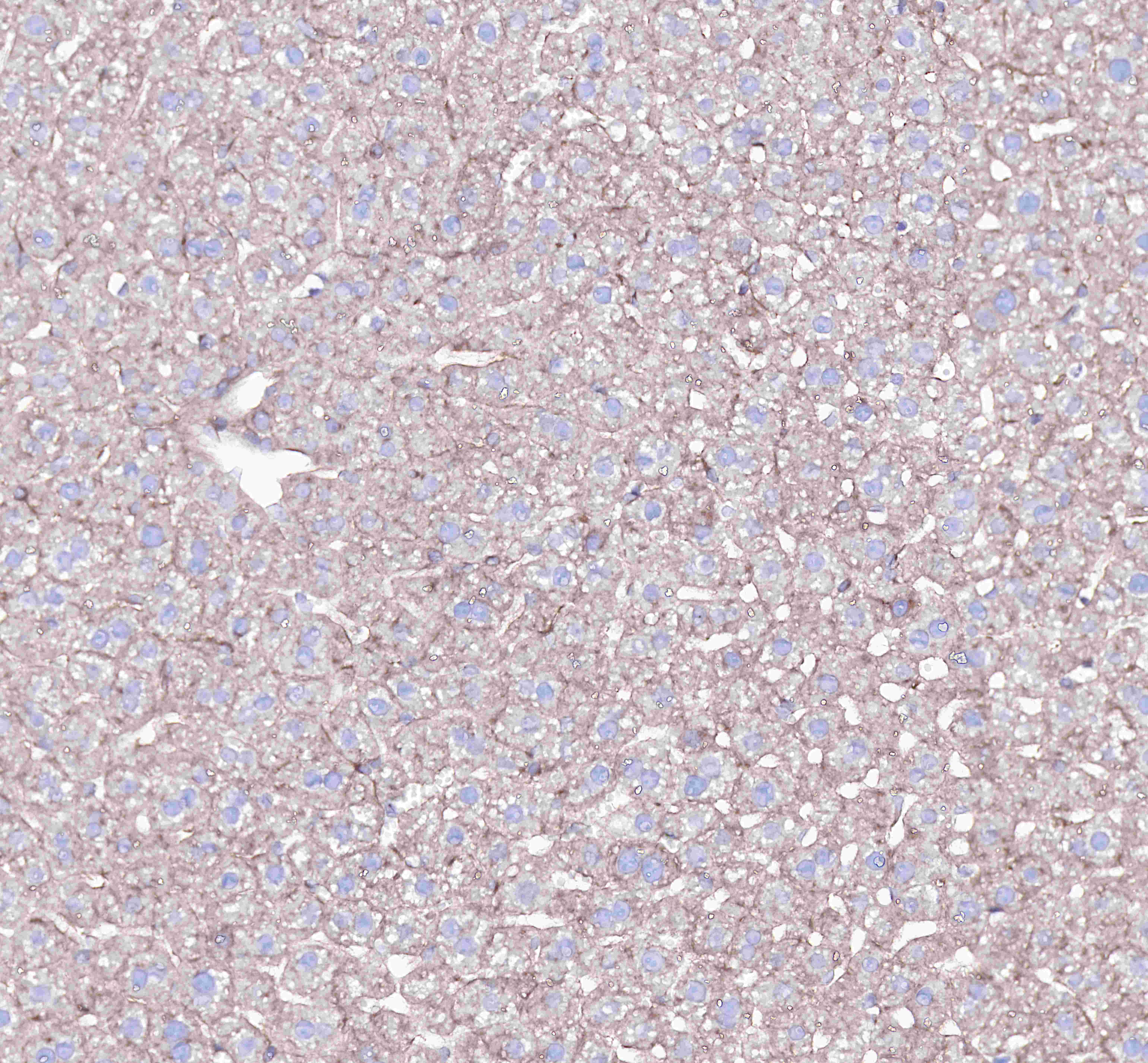
IHC shows positive staining in paraffin-embedded mouse liver.
Anti-β-tubulin antibody was used at 1/1000 dilution, followed by a Goat Anti-Rabbit IgG H&L (HRP) ready to use.
Counterstained with hematoxylin.
Heat mediated antigen retrieval with Tris/EDTA buffer pH9.0 was performed before commencing with IHC staining protocol.
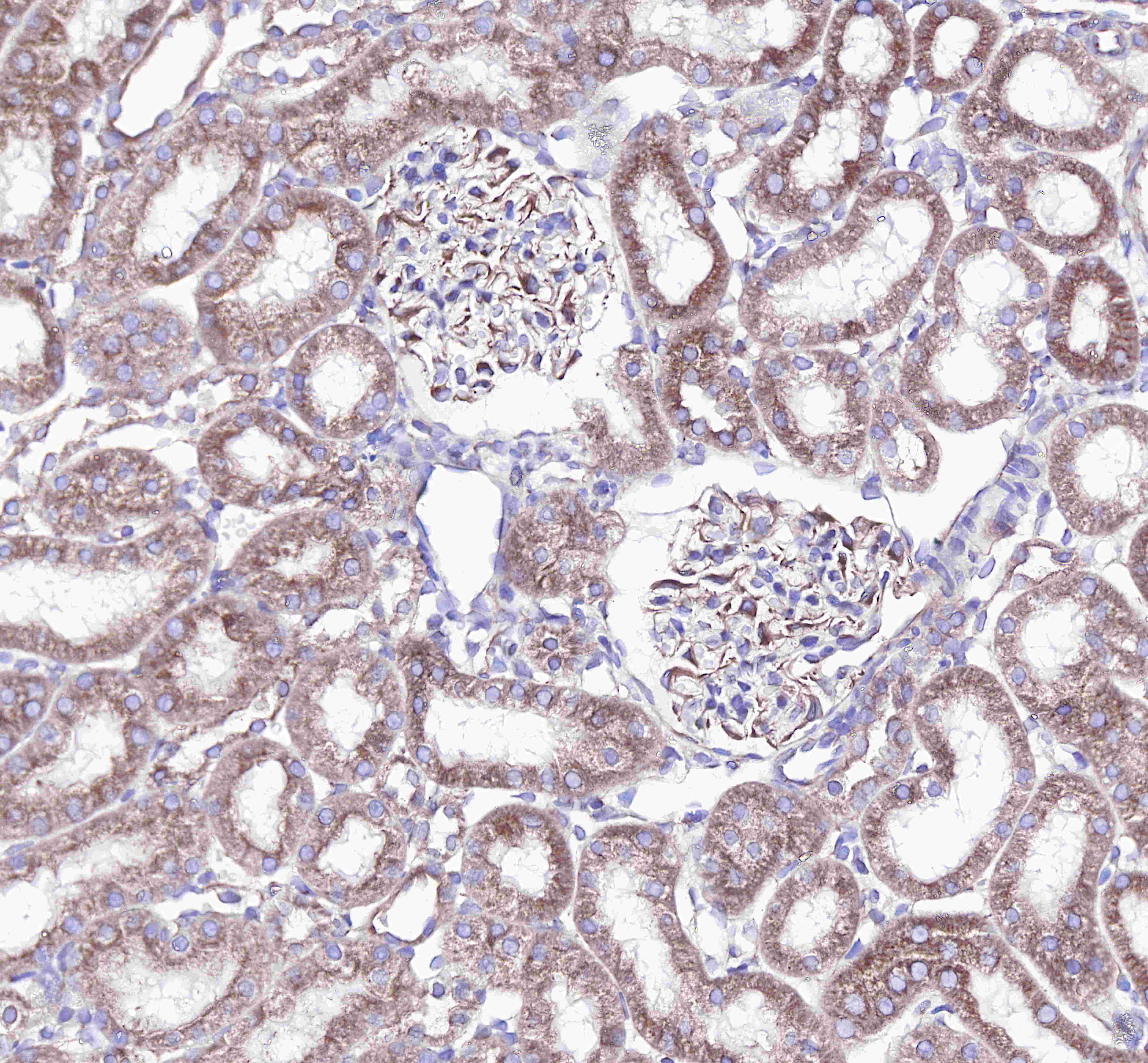
IHC shows positive staining in paraffin-embedded rat kidney.
Anti-β-tubulin antibody was used at 1/1000 dilution, followed by a Goat Anti-Rabbit IgG H&L (HRP) ready to use.
Counterstained with hematoxylin.
Heat mediated antigen retrieval with Tris/EDTA buffer pH9.0 was performed before commencing with IHC staining protocol.
免疫细胞化学
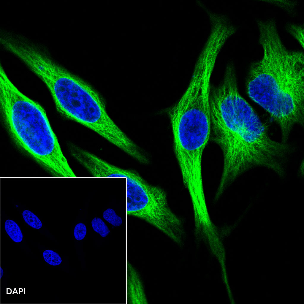
ICC shows positive staining in HeLa cells. Anti-β-tubulin antibody was used at 1/500 dilution (Green) and incubated overnight at 4°C. Goat polyclonal Antibody to Rabbit IgG - H&L (Alexa Fluor® 488) was used as secondary antibody at 1/1000 dilution. The cells were fixed with 4% PFA and permeabilized with 0.1% PBS-Triton X-100. Nuclei were counterstained with DAPI (Blue).


评论(0)