


 大包装询价
大包装询价 产品介绍 评论(0)
宿主来源
Rabbit抗原名称
HSP60分子别名
60 kDa chaperonin; Chaperonin 60 (CPN60); HuCHA60; Mitochondrial matrix protein P1; P60 lymphocyte protein免疫原
Recombinant Protein细胞定位
IntracellularAccession
P10809克隆号
SDT-R012抗体类型
Rabbit mAb应用
ICFCM, IHC-P, ICC, WB反应种属 ?
Hu, Ms, Rt纯化方式
Protein A浓度
0.5mg/ml分子量
60kDa
标记
Unconjugated性状
Liquid缓冲体系
PBS, 40% Glycerol, 0.05%BSA, 0.03% Proclin 300
储存条件
12 months from date of receipt / reconstitution, -20 °C as supplied.
| 应用 | 稀释度 |
|---|---|
| ICC | 1:500 |
| IHC-P | 1:500 |
| ICFCM | 1:500 |
| WB | 1:1000-1:20000 |
HSP60, also known as chaperonins (Cpn), is a family of heat shock proteins originally sorted by their 60kDa molecular mass. They prevent misfolding of proteins during stressful situations such as high heat, by assisting protein folding. HSP60 belongs to a large class of molecules that assist protein folding, called molecular chaperones.
免疫印迹
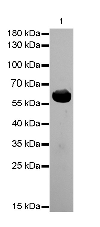
WB result of Hsp60 Rabbit mAb
Primary antibody: Hsp60 Rabbit mAb at 1/1000 dilution
Lane 1: Hela whole cell lysate 20 µg
Secondary antibody: Goat Anti-Rabbit IgG, (H+L), HRP conjugated at 1/10000 dilution
Predicted MW: 60 kDa
Observed MW: 60 kDa
Exposure time: 3 seconds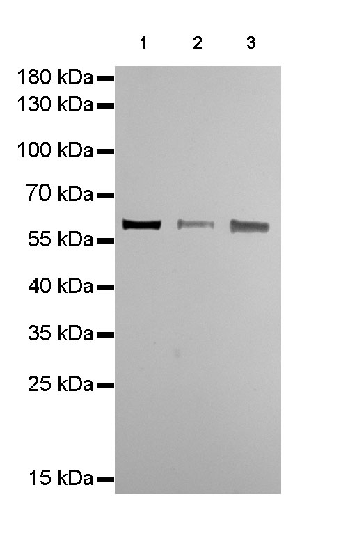
WB result of Hsp60 Rabbit mAb
Primary antibody: Hsp60 Rabbit mAb at 1/1000 dilution
Lane 1: mouse heart lysate 20 µg
Lane 2: mouse spleen lysate 20 µg
Lane 3: NIH/3T3 whole cell lysate 20 µg
Secondary antibody: Goat Anti-Rabbit IgG, (H+L), HRP conjugated at 1/10000 dilution
Predicted MW: 60 kDa
Observed MW: 60 kDa
Exposure time: 3 seconds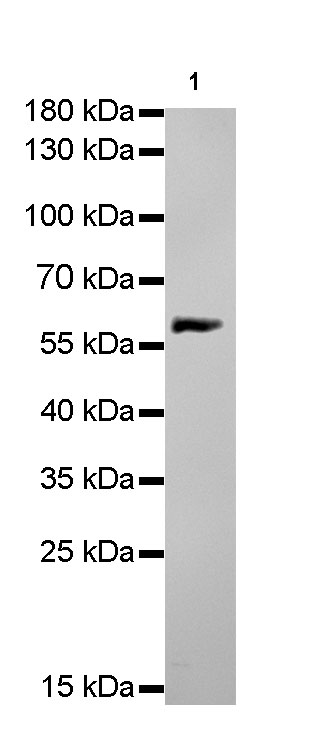
WB result of Hsp60 Rabbit mAb
Primary antibody: Hsp60 Rabbit mAb at 1/1000 dilution
Lane 1: rat brain lysate 20 µg
Secondary antibody: Goat Anti-Rabbit IgG, (H+L), HRP conjugated at 1/10000 dilution
Predicted MW: 60 kDa
Observed MW: 60 kDa
Exposure time: 15 seconds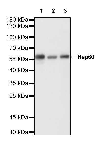
WB result of Hsp60 Rabbit mAb
Primary antibody: Hsp60 Rabbit mAb at 1/20000 dilution
Lane 1: HeLa whole cell lysate 20 µg
Lane 2: NIH/3T3 whole cell lysate 20 µg
Lane 3: rat heart lysate 20 µg
Secondary antibody: Goat Anti-Rabbit IgG, (H+L), HRP conjugated at 1/10000 dilution
Predicted MW: 60 kDa
Observed MW: 60 kDa
Exposure time: 120 s
流式分析
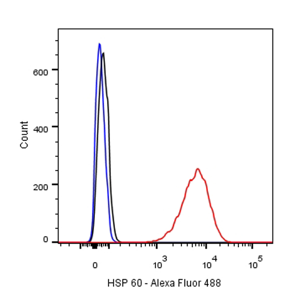
Flow cytometric analysis of HeLa cells labelling HSP 60 antibody at 1/500 (0.1ug) dilution/ (red) compared with a Rabbit monoclonal IgG (Black) isotype control and an unlabelled control (cells without incubation with primary antibody and secondary antibody) (Blue). Goat Anti-Rabbit IgG Alexa Fluor® 488 was used as the secondary antibody.
免疫组化
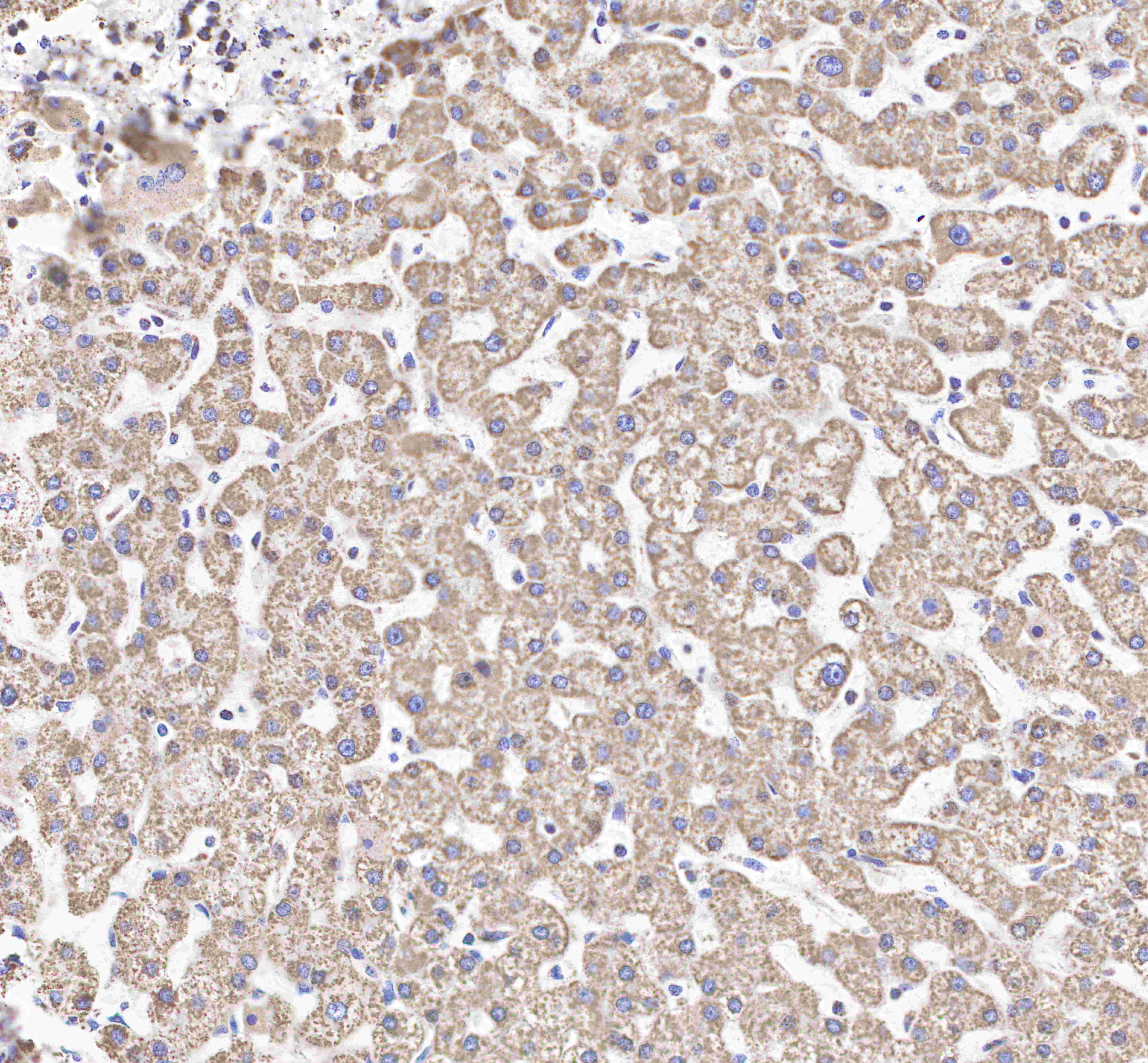
IHC shows positive staining in paraffin-embedded human liver.
Anti-Hsp60 antibody was used at 1/500 dilution, followed by a Goat Anti-Rabbit IgG H&L (HRP) ready to use.
Counterstained with hematoxylin.
Heat mediated antigen retrieval with Tris/EDTA buffer pH9.0 was performed before commencing with IHC staining protocol.
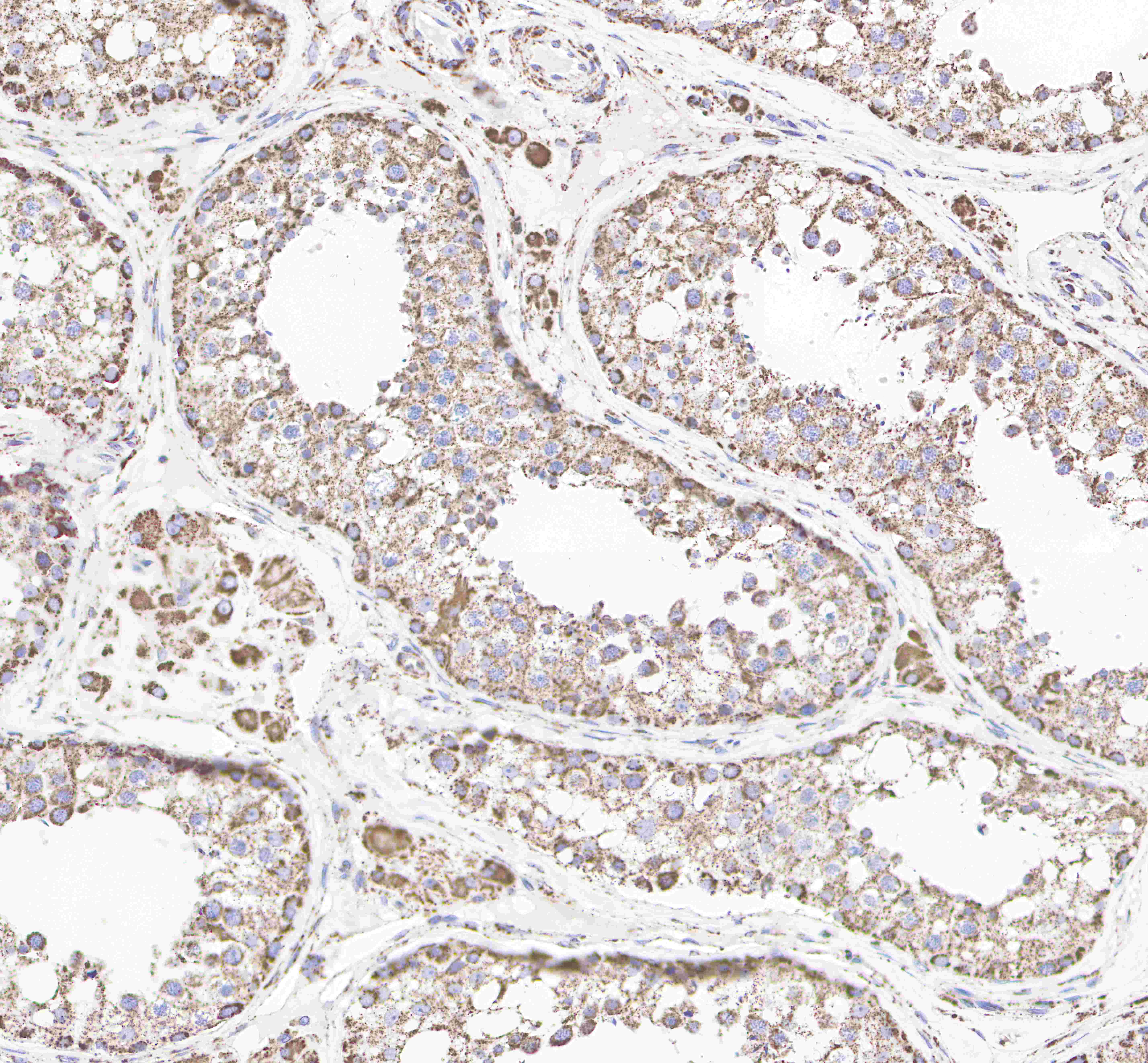
IHC shows positive staining in paraffin-embedded human testis.
Anti-Hsp60 antibody was used at 1/500 dilution, followed by a Goat Anti-Rabbit IgG H&L (HRP) ready to use.
Counterstained with hematoxylin.
Heat mediated antigen retrieval with Tris/EDTA buffer pH9.0 was performed before commencing with IHC staining protocol.
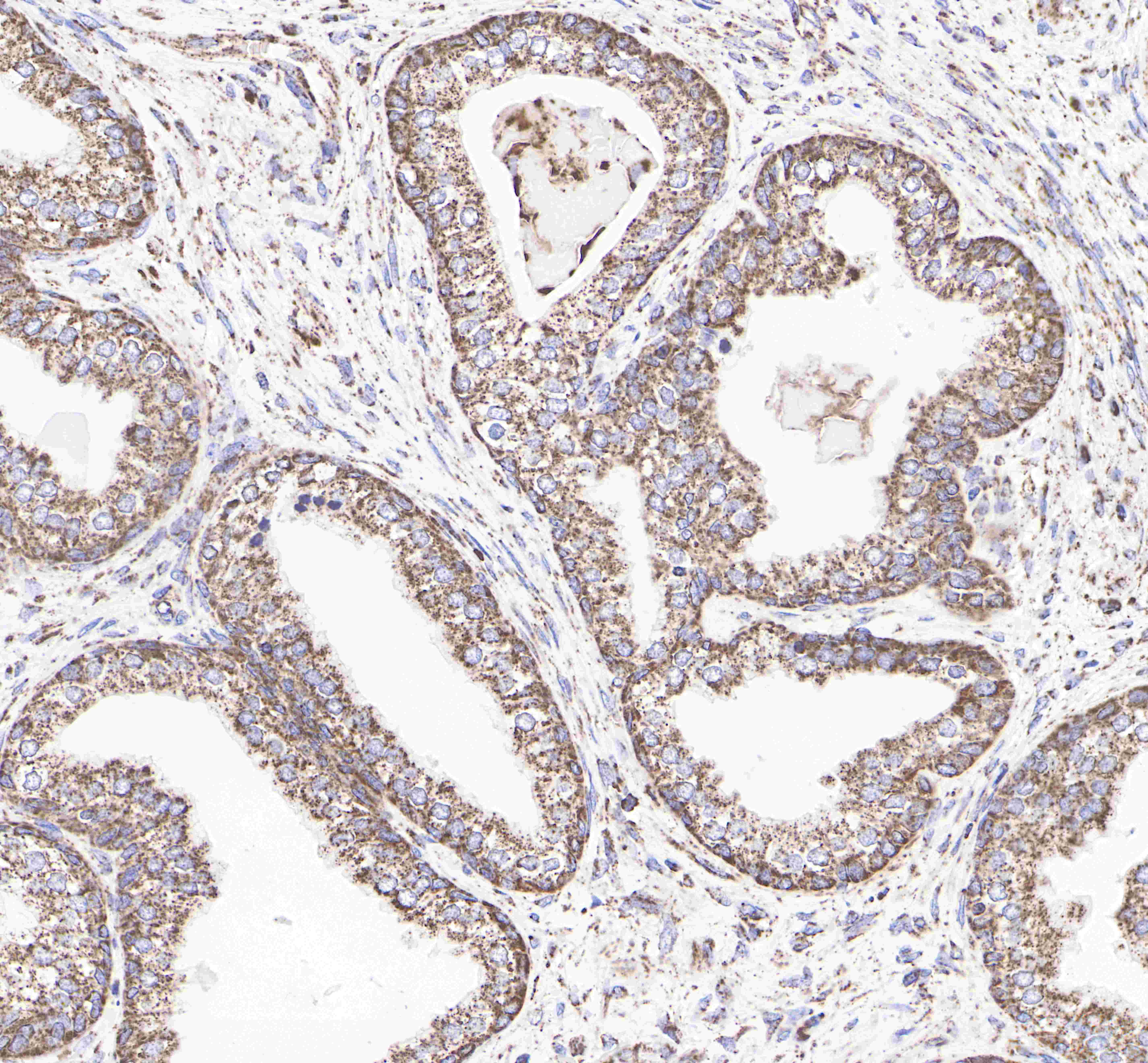
IHC shows positive staining in paraffin-embedded human prostate.
Anti-Hsp60 antibody was used at 1/500 dilution, followed by a Goat Anti-Rabbit IgG H&L (HRP) ready to use.
Counterstained with hematoxylin.
Heat mediated antigen retrieval with Tris/EDTA buffer pH9.0 was performed before commencing with IHC staining protocol.
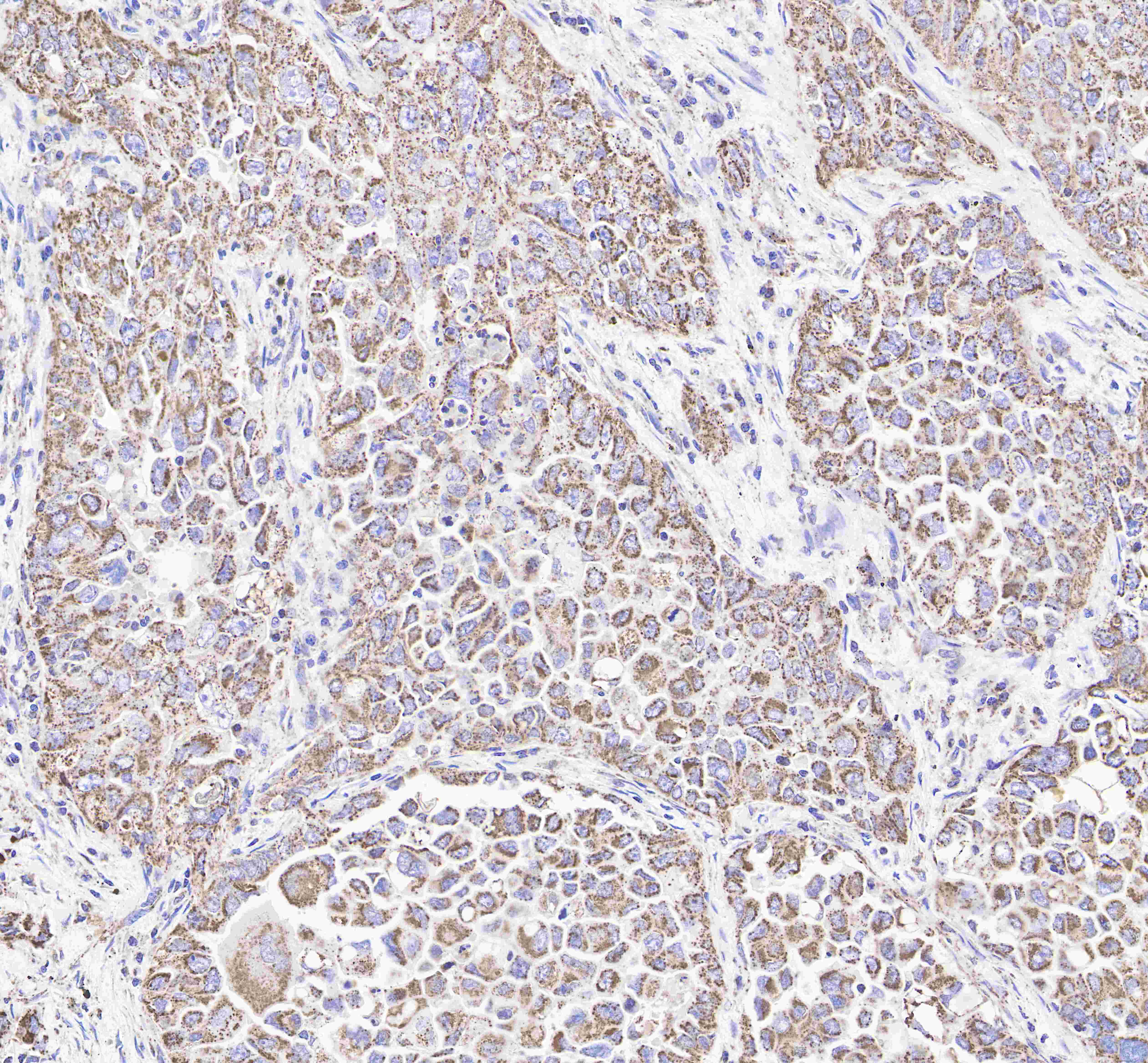
IHC shows positive staining in paraffin-embedded human lung cancer.
Anti-Hsp60 antibody was used at 1/500 dilution, followed by a Goat Anti-Rabbit IgG H&L (HRP) ready to use.
Counterstained with hematoxylin.
Heat mediated antigen retrieval with Tris/EDTA buffer pH9.0 was performed before commencing with IHC staining protocol.
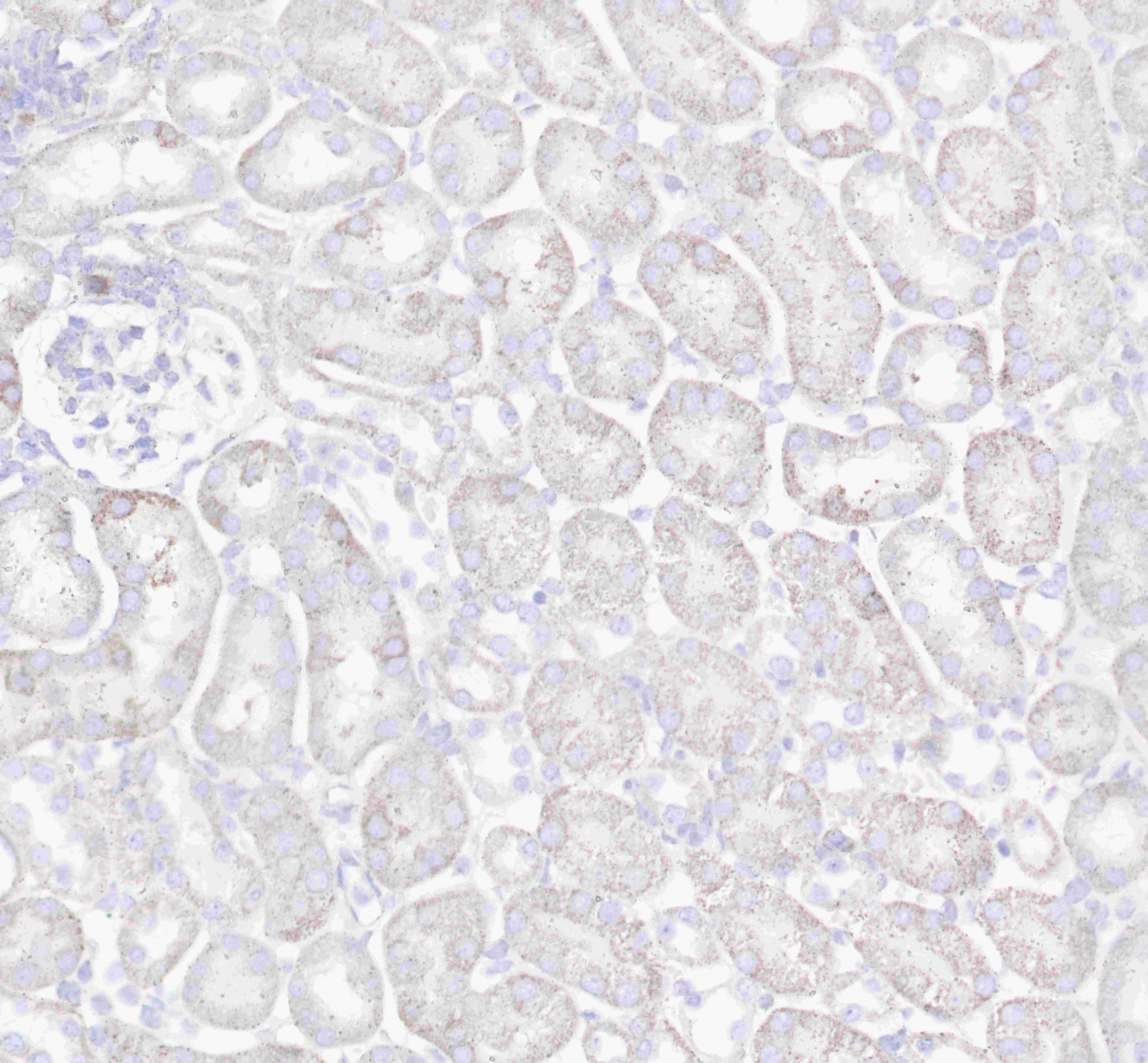
IHC shows positive staining in paraffin-embedded mouse kidney.
Anti-Hsp60 antibody was used at 1/500 dilution, followed by a Goat Anti-Rabbit IgG H&L (HRP) ready to use.
Counterstained with hematoxylin.
Heat mediated antigen retrieval with Tris/EDTA buffer pH9.0 was performed before commencing with IHC staining protocol.
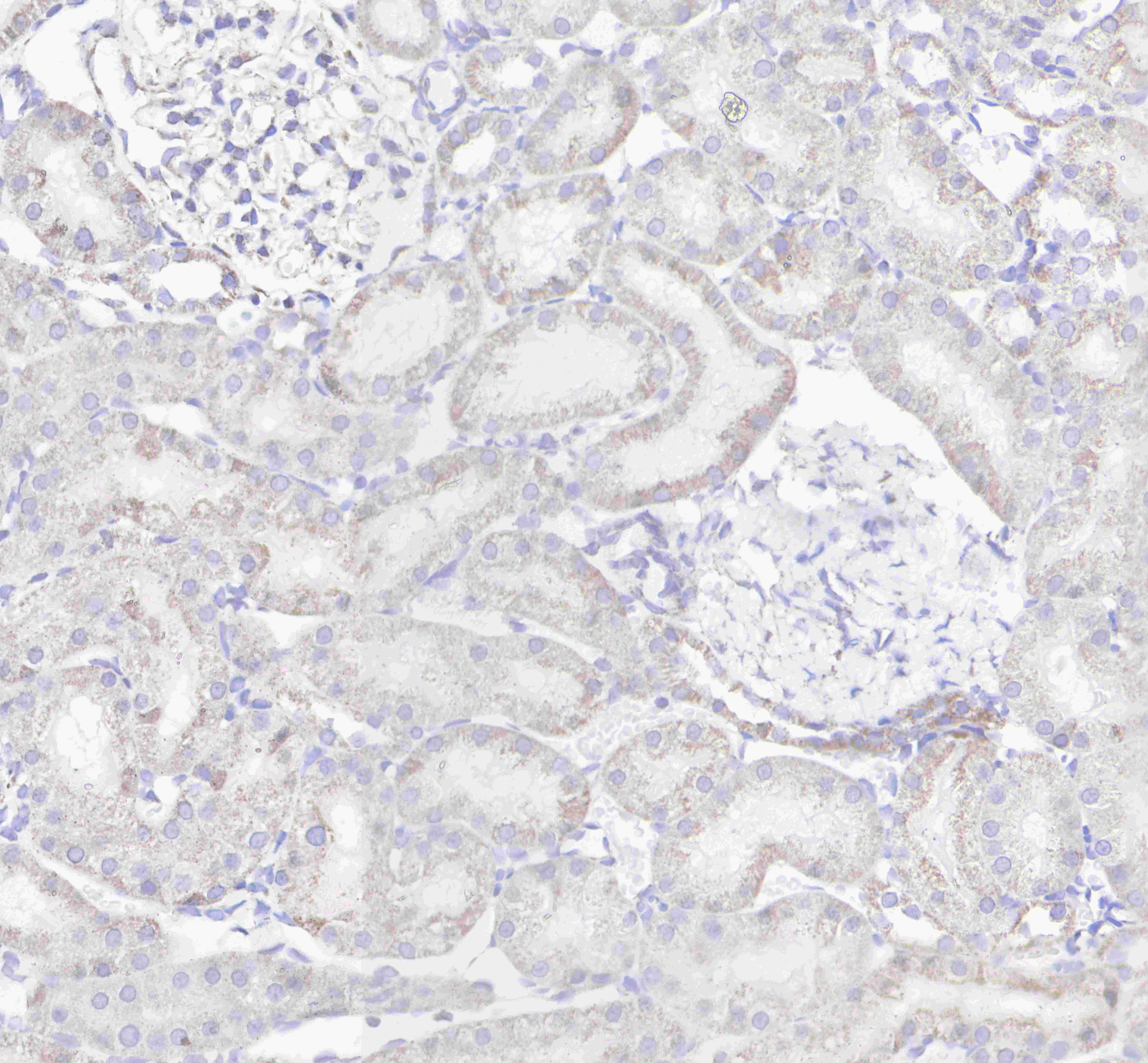
IHC shows positive staining in paraffin-embedded rat kidney.
Anti-Hsp60 antibody was used at 1/500 dilution, followed by a Goat Anti-Rabbit IgG H&L (HRP) ready to use.
Counterstained with hematoxylin.
Heat mediated antigen retrieval with Tris/EDTA buffer pH9.0 was performed before commencing with IHC staining protocol.
免疫细胞化学
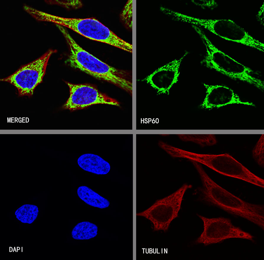
ICC shows positive staining in HeLa cells. Anti-HSP60 antibody was used at 1/500 dilution (Green) and incubated overnight at 4°C. Goat polyclonal Antibody to Rabbit IgG - H&L (Alexa Fluor® 488) was used as secondary antibody at 1/1000 dilution. The cells were fixed with 4%PFA and permeabilized with 0.1% PBS-Triton X-100. Nuclei were counterstained with DAPI (Blue). Counterstain with tubulin (red).

评论(0)