


 大包装询价
大包装询价 产品介绍 评论(0)
宿主来源
Rabbit抗原名称
ERG分子别名
Transcriptional regulator ERG, Transforming protein ERG免疫原
Synthetic Peptide细胞定位
Nucleus, CytoplasmAccession
P11308克隆号
SDT-190-65抗体类型
Rabbit mAb应用
ICFCM, IHC-P, ICC, WB反应种属 ?
Hu纯化方式
Protein A浓度
1 mg/ml性状
Liquid缓冲体系
PBS
储存条件
12 months from date of receipt, 4°C as supplied
| 应用 | 稀释度 |
|---|---|
| WB | 1:500 |
| IHC-P | 1:1000 |
| ICFCM | 1:250 |
| ICC | 1:250 |
ERG is an ETS family transcription factor (TF) frequently involved in human cancers, including leukemia [PMID: 21664289], prostate cancer [PMID: 16254181] and Ewing’s sarcoma [PMID: 8223458, PMID: 10561219]. It functions as an oncogene through chromosomal translocations or overexpression. In human leukemia, ERG was initially thought to play a role in leukemogenesis based on the t(16;21) translocation in acute myeloid leukemia (AML), leading to formation of the FUS (TLS)-ERG fusion [PMID: 8234289]. In some cases of AML with this translocation, the leukemia exhibits features of acute megakaryoblastic leukemia [PMID: 12091356].
免疫印迹
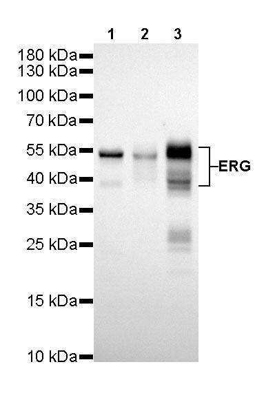
WB result of ERG Rabbit mAb
Primary antibody: ERG Rabbit mAb at 1/500 dilution
Lane 1: Jurkat whole cell lysate 20 µg
Lane 2: Hela whole cell lysate 20 µg
Lane 3: MOLT-4 whole cell lysate 20 µg
Secondary antibody: Goat Anti-Rabbit IgG, (H+L), HRP conjugated at 1/10000 dilution
Predicted MW: 41~55 kDa
Observed MW: 41~55 kDa
Exposure time: 180s
流式分析
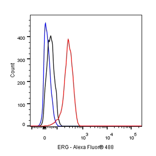
Flow cytometric analysis of 4% PFA fixed 90% methanol permeabilized THP-1 (Human monocytic leukemia monocyte) cells labelling ERG antibody at 1/250 (0.1 μg) dilution / (red) compared with a Rabbit monoclonal IgG (Black) isotype control and an unlabelled control (cells without incubation with primary antibody and secondary antibody) (Blue). Goat Anti - Rabbit IgG Alexa Fluor® 488 was used as the secondary antibody.
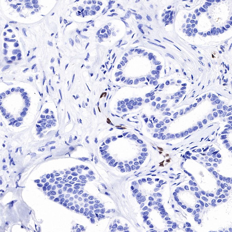
免疫组化
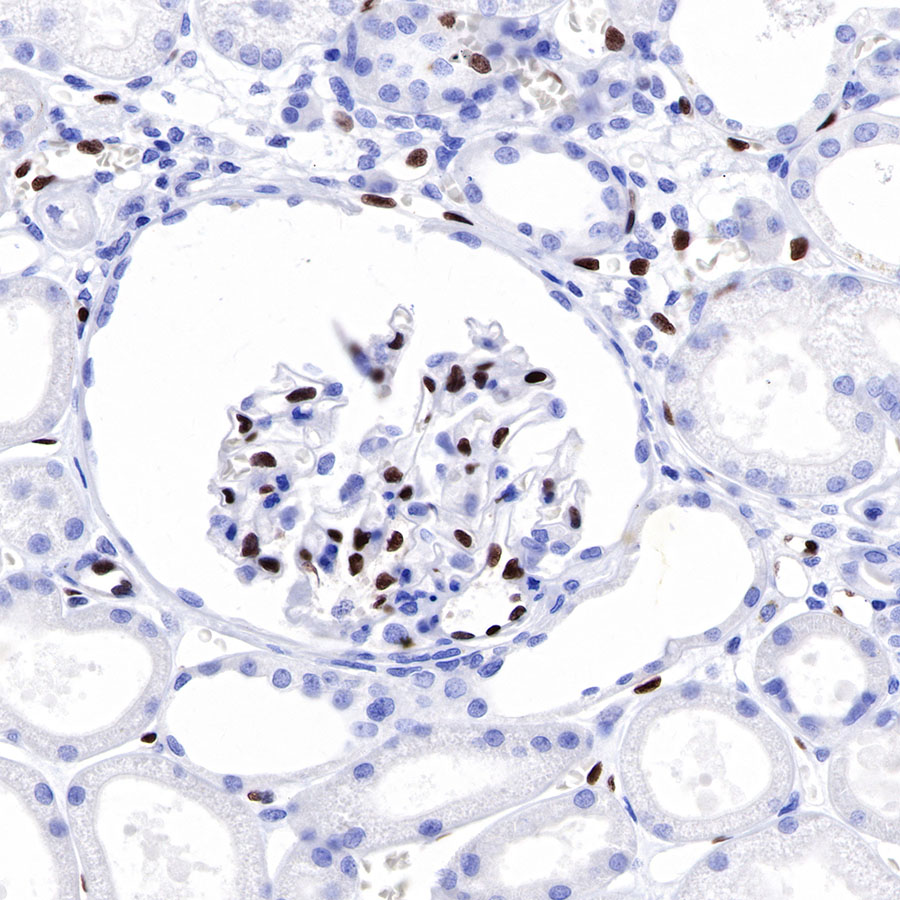
IHC shows positive staining in paraffin-embedded human kidney. Anti-ERG antibody was used at 1/1000 dilution, followed by a HRP Polymer for Mouse & Rabbit IgG (ready to use). Counterstained with hematoxylin. Heat mediated antigen retrieval with Tris/EDTA buffer pH9.0 was performed before commencing with IHC staining protocol.
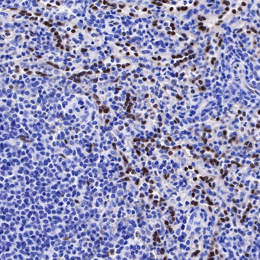
IHC shows positive staining in paraffin-embedded human spleen. Anti-ERG antibody was used at 1/1000 dilution, followed by a HRP Polymer for Mouse & Rabbit IgG (ready to use). Counterstained with hematoxylin. Heat mediated antigen retrieval with Tris/EDTA buffer pH9.0 was performed before commencing with IHC staining protocol.
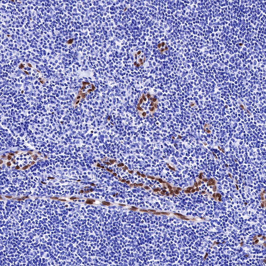
IHC shows positive staining in paraffin-embedded human tonsil. Anti-ERG antibody was used at 1/1000 dilution, followed by a HRP Polymer for Mouse & Rabbit IgG (ready to use). Counterstained with hematoxylin. Heat mediated antigen retrieval with Tris/EDTA buffer pH9.0 was performed before commencing with IHC staining protocol.
IHC shows positive staining in paraffin-embedded human breast cancer. Anti-ERG antibody was used at 1/1000 dilution, followed by a HRP Polymer for Mouse & Rabbit IgG (ready to use). Counterstained with hematoxylin. Heat mediated antigen retrieval with Tris/EDTA buffer pH9.0 was performed before commencing with IHC staining protocol.
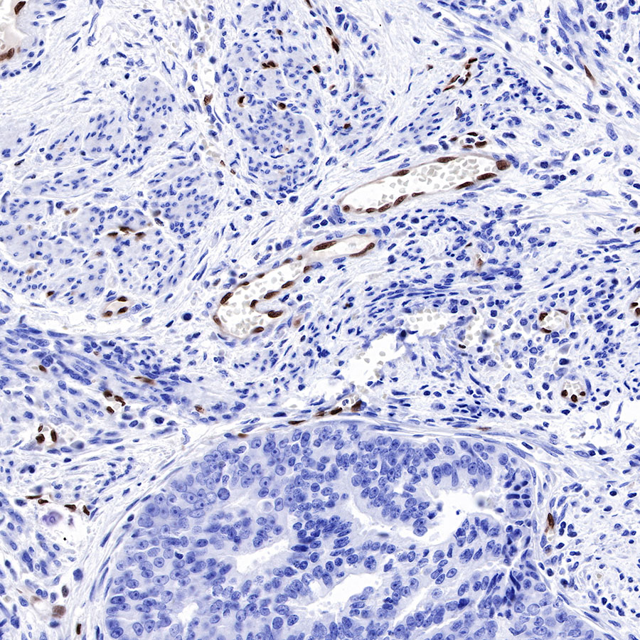
IHC shows positive staining in paraffin-embedded human endometrial cancer. Anti-ERG antibody was used at 1/1000 dilution, followed by a HRP Polymer for Mouse & Rabbit IgG (ready to use). Counterstained with hematoxylin. Heat mediated antigen retrieval with Tris/EDTA buffer pH9.0 was performed before commencing with IHC staining protocol.
免疫细胞化学
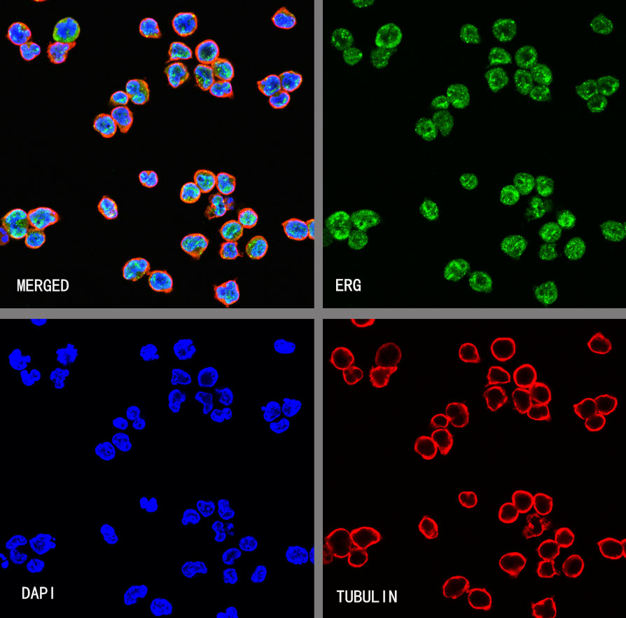
ICC shows positive staining in Jurkat cells. Anti-ERG antibody was used at 1/100 dilution (Green) and incubated overnight at 4°C. Goat polyclonal Antibody to Rabbit IgG - H&L (Alexa Fluor® 488) was used as secondary antibody at 1/1000 dilution. The cells were fixed with 4% PFA and permeabilized with 0.1% PBS-Triton X-100. Nuclei were counterstained with DAPI (Blue). Counterstain with tubulin (Red).


评论(0)