


 大包装询价
大包装询价 产品介绍 评论(0)
宿主来源
Rabbit抗原名称
CD22分子别名
BL-CAM, Siglec-2, T-cell surface antigen Leu-14免疫原
N/A细胞定位
Cell membraneAccession
P20273克隆号
SDT-R113抗体类型
Rabbit mAb应用
IHC-P, ICC, WB, IP反应种属 ?
Hu纯化方式
Protein A浓度
1 mg/ml性状
Liquid缓冲体系
PBS储存条件
12 months from date of receipt, 4°C as supplied
| 应用 | 稀释度 |
|---|---|
| WB | 1:1000 |
| IHC-P | 1:1000 |
| ICC | 1:500 |
| IP | 1:50 |
CD22 and CD19 represent two specialized costimulatory or coreceptor cell surface molecules [PMID: 9476671, PMID: 9175829, PMID: 15364057, PMID: 9203413, PMID: 9047233] that also function as “response regulators” to modulate the intensity, quality, and duration of homeostatic and BCR‐induced signals [PMID: 9695183, PMID: 9695185]. Response regulators carry out broader functions than costimulatory molecules because they establish intrinsic signaling thresholds that provide a context for other transmembrane and cytoplasmic signals. CD22 is also a lectin‐like member of the Ig superfamily expressed exclusively by all mature B‐lineage cells, which binds ligands in vivo to regulate BCR and CD19 signal transduction, and provide essential survival signals.
免疫印迹
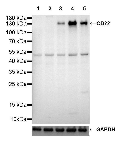
WB result of CD22 Rabbit mAb
Primary antibody: CD22 Rabbit mAb at 1/1000 dilution
Lane 1: K562 whole cell lysate 20 µg
Lane 2: Jurkat whole cell lysate 20 µg
Lane 3: Raji whole cell lysate 20 µg
Lane 4: Daudi whole cell lysate 20 µg
Lane 5: Ramos whole cell lysate 20 µg
Negative control: K562 whole cell lysate
Negative control: Jurkat whole cell lysate
Secondary antibody: Goat Anti-Rabbit IgG, (H+L), HRP conjugated at 1/10000 dilution
Predicted MW: 140 kDa
Observed MW: 140 kDa
免疫沉淀
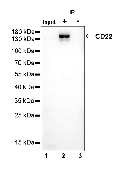
CD22 Rabbit mAb at 1/50 dilution (1 µg) immunoprecipitating CD22 in 0.4 mg Daudi whole cell lysate.
Western blot was performed on the immunoprecipitate using CD22 Rabbit mAb at 1/1000 dilution.
Secondary antibody (HRP) for IP was used at 1/400 dilution.
Lane 1: Daudi whole cell lysate 10 µg (Input)
Lane 2: CD22 Rabbit mAb IP in Daudi whole cell lysate
Lane 3: Rabbit monoclonal IgG IP in Daudi whole cell lysate
Predicted MW: 140 kDa
Observed MW: 140 kDa
免疫组化
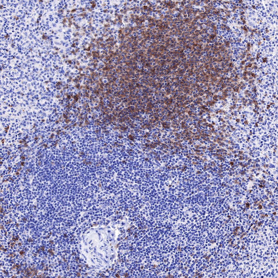
IHC shows positive staining in paraffin-embedded human spleen. Anti-CD22 antibody was used at 1/1000 dilution, followed by a HRP Polymer for Mouse & Rabbit IgG (ready to use). Counterstained with hematoxylin. Heat mediated antigen retrieval with Tris/EDTA buffer pH9.0 was performed before commencing with IHC staining protocol.
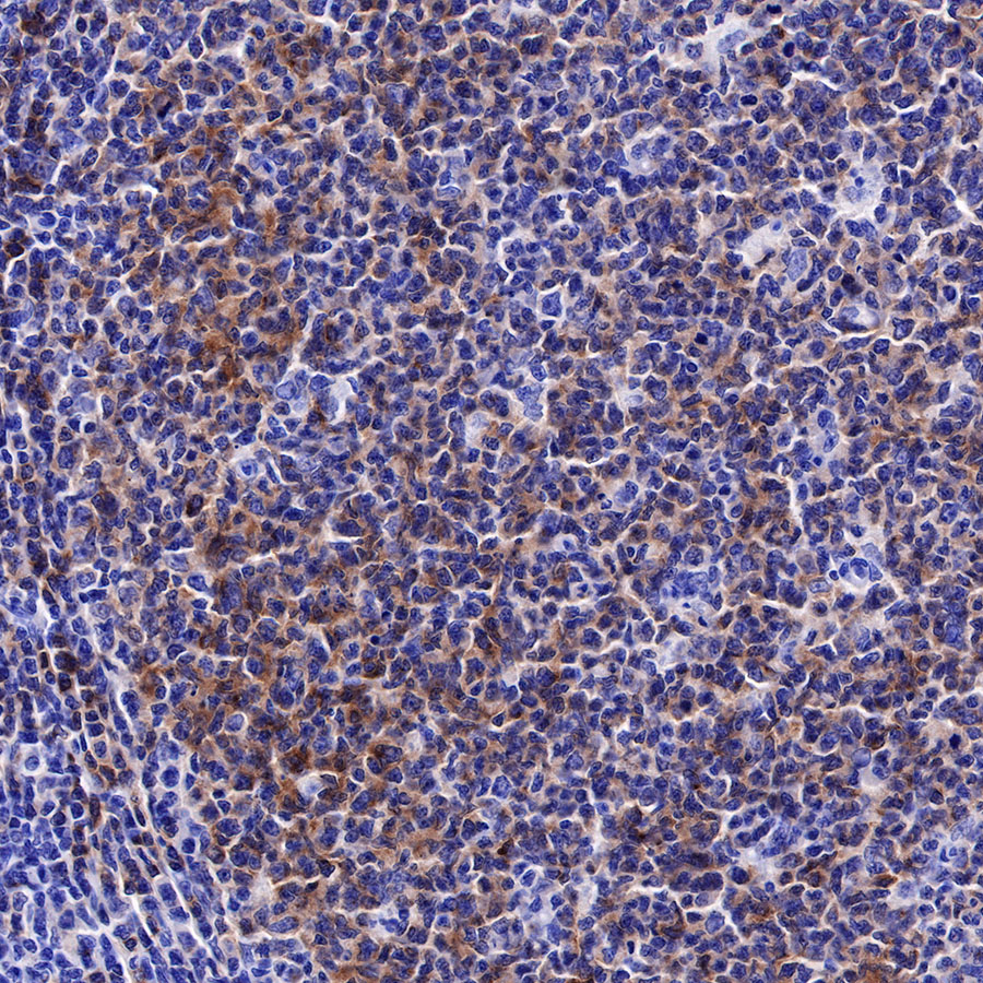
IHC shows positive staining in paraffin-embedded human tonsil. Anti-CD22 antibody was used at 1/1000 dilution, followed by a HRP Polymer for Mouse & Rabbit IgG (ready to use). Counterstained with hematoxylin. Heat mediated antigen retrieval with Tris/EDTA buffer pH9.0 was performed before commencing with IHC staining protocol.
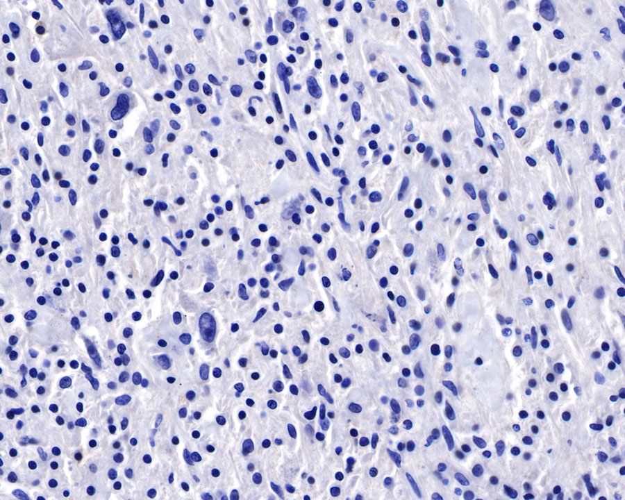
Negative control: IHC shows negative staining in paraffin-embedded human Hodgkin's lymphoma. Anti-CD22 antibody was used at 1/1000 dilution, followed by a HRP Polymer for Mouse & Rabbit IgG (ready to use). Counterstained with hematoxylin. Heat mediated antigen retrieval with Tris/EDTA buffer pH9.0 was performed before commencing with IHC staining protocol.
免疫细胞化学
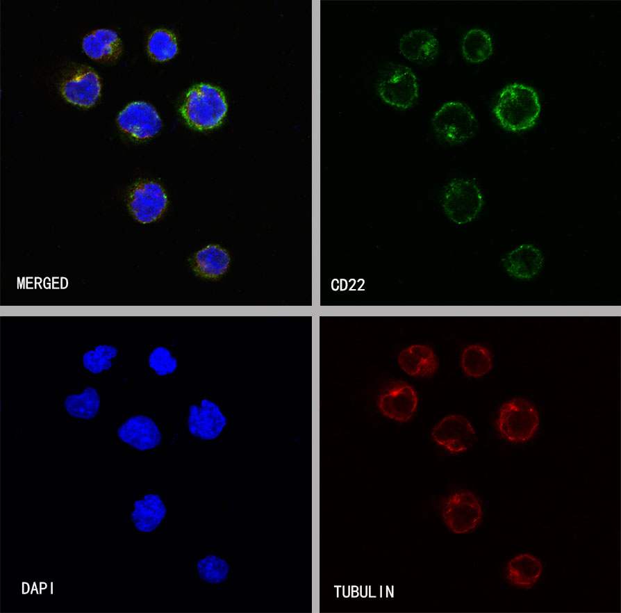
ICC shows positive staining in Daudi cells. Anti-CD22 antibody was used at 1/500 dilution (Green) and incubated overnight at 4°C. Goat polyclonal Antibody to Rabbit IgG - H&L (Alexa Fluor® 488) was used as secondary antibody at 1/1000 dilution. The cells were fixed with 4% PFA and permeabilized with 0.1% PBS-Triton X-100. Nuclei were counterstained with DAPI (Blue). Counterstain with tubulin (red).


评论(0)