


 大包装询价
大包装询价 产品介绍 评论(0)
宿主来源
Rabbit抗原名称
CD40分子别名
B-cell surface antigen CD40,Bp50 ,CD40L receptor,CDw40免疫原
Recombinant Protein细胞定位
Cell membrane, SecretedAccession
P25942克隆号
SDT-145-45抗体类型
Rabbit mAb应用
IHC-P, FCM, WB, IP反应种属 ?
Hu纯化方式
Protein A浓度
0.5mg/ml性状
Liquid缓冲体系
PBS, 40% Glycerol, 0.05%BSA, 0.03% Proclin 300储存条件
12 months from date of receipt / reconstitution, -20 °C as supplied
| 应用 | 稀释度 |
|---|---|
| FCM | 1:500 |
| IHC-P | 1:100-500 |
| IP | 1:25 |
| WB | 1:1000 |
- The CD40 is a type I transgender protein that is transmitted in the immune system's B cells and special antigen to the surface of the cells and many non -hematopoietic cell types and cancer cells. It belongs to the TNF receptor super family.CD40 is expressed in B cells, foam dendritic cells, dendritic cells, activated monocytes, macrophages, endothelial cells, vascular smooth muscle cells, and several tumor cell lines. CD40 interacts with CD40 Ligand (CD40L/TNFSF5).CD40/CD40L is essential for the beginning and progress of cells and body fluid adaptive immunity, and it is an important area of concern in the study of tumor immunology, neurodegeneration, vascular disease and inflammatory diseases.
验证数据
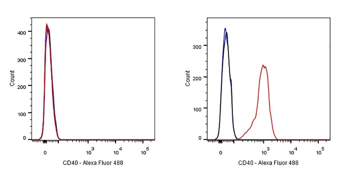
Flow cytometric analysis of Jurkat (left) / Ramos (right) cells labelling CD40 antibody at 1/500 dilution (0.1ug)/ (red) compared with a Rabbit monoclonal IgG (Black) isotype control and an unlabelled control (cells without incubation with primary antibody and secondary antibody) (Blue). Goat Anti-Rabbit IgG Alexa Fluor® 488 was used as the secondary antibody. Negative control: Jurkat
免疫印迹
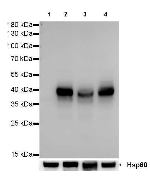
WB result of CD40 Rabbit mAb
Primary antibody:CD40 Rabbit mAb at 1/1000 dilution
Lane 1: Jurkat whole cell lysate 20 µg
Lane 2: Raji whole cell lysate 20 µg
Lane 3: Ramos whole cell lysate 20 µg
Lane 4: Daudi whole cell lysate 20 µg
Negative control: Jurkat whole cell lysateSecondary antibody: Goat Anti-Rabbit IgG, (H+L), HRP conjugated at 1/10000 dilution
Predicted MW: 42 kDa
Observed MW: 39 kDa
免疫沉淀
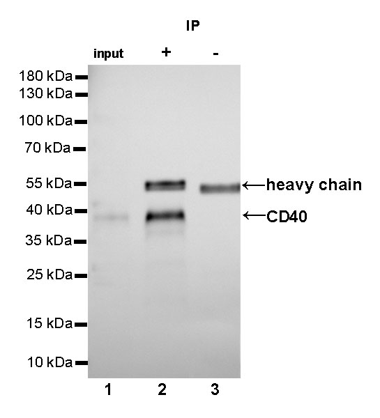
CD40 Rabbit mAb at 1/25 dilution (2µg) immunoprecipitating CD40 in 0.4mg Raji whole cell lysate.
Western blot was performed on the immunoprecipitate using CD40 Rabbit mAb at 1/1000 dilution.
Secondary antibody (HRP) for IP was used at 1/400 dilution.
Lane 1: Raji whole cell lysate 10µg (input)
Lane 2 (+): CD40 Rabbit mAb IP in Raji whole cell lysate
Lane 3 (-): Rabbit monoclonal IgG IP in Raji whole cell lysate
Predicted MW: 42 kDa
Observed MW: 39 kDa
免疫组化

IHC shows positive staining in paraffin-embedded human tonsil.
Anti-CD40 antibody was used at 1/100 dilution, followed by a Goat Anti-Rabbit IgG H&L (HRP) ready to use. Counterstained with hematoxylin.
Heat mediated antigen retrieval with Tris/EDTA buffer pH9.0 was performed before commencing with IHC staining protocol.
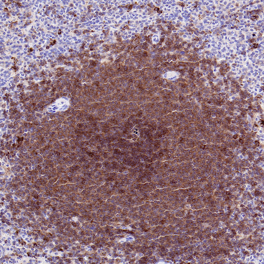
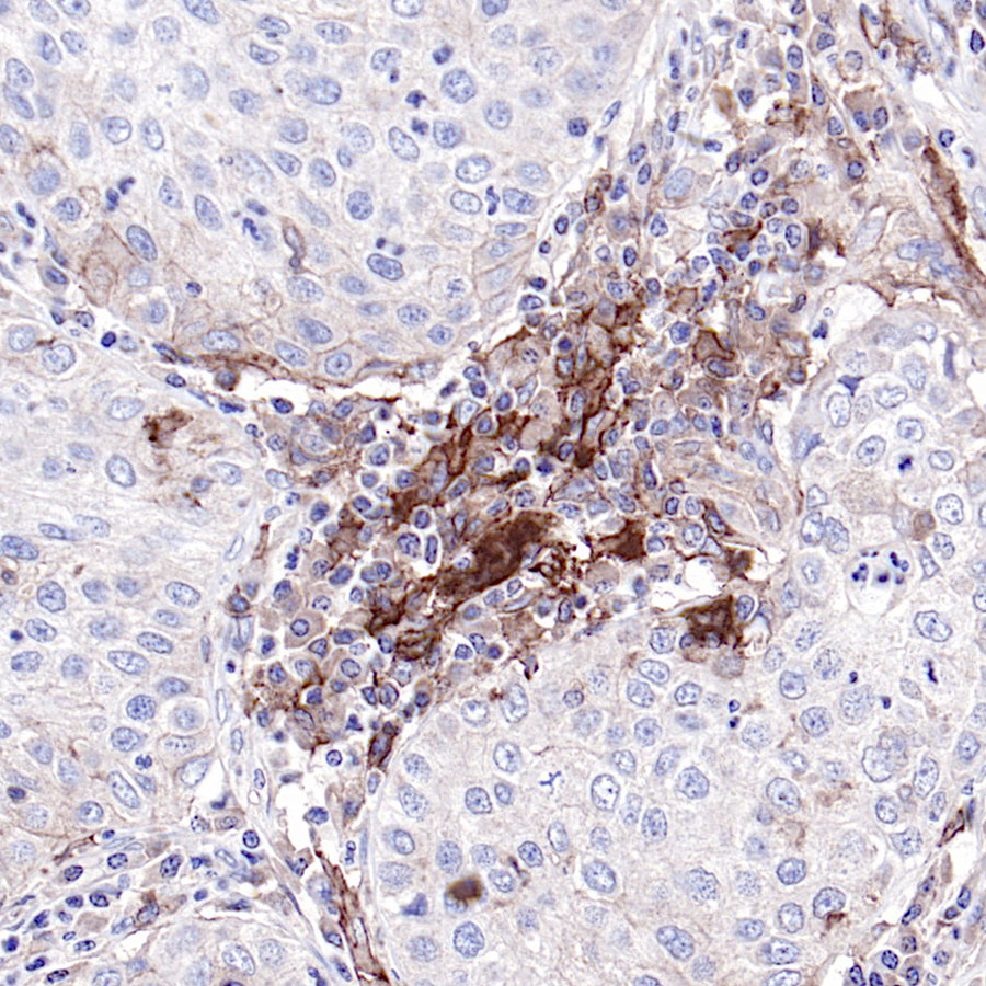
IHC shows positive staining in paraffin-embedded human spleen.
Anti-CD40 antibody was used at 1/100 dilution, followed by a Goat Anti-Rabbit IgG H&L (HRP) ready to use. Counterstained with hematoxylin.
Heat mediated antigen retrieval with Tris/EDTA buffer pH9.0 was performed before commencing with IHC staining protocol.
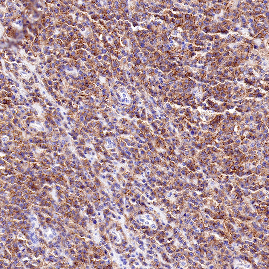
IHC shows positive staining in paraffin-embedded human diffuse large B-cell lymphoma.
Anti-CD40 antibody was used at 1/500 dilution, followed by a Goat Anti-Rabbit IgG H&L (HRP) ready to use. Counterstained with hematoxylin.
Heat mediated antigen retrieval with Tris/EDTA buffer pH9.0 was performed before commencing with IHC staining protocol.
IHC shows positive staining in paraffin-embedded human lung squamous cancer.
Anti-CD40 antibody was used at 1/100 dilution, followed by a Goat Anti-Rabbit IgG H&L (HRP) ready to use. Counterstained with hematoxylin.
Heat mediated antigen retrieval with Tris/EDTA buffer pH9.0 was performed before commencing with IHC staining protocol.


评论(0)