


CD3d Recombinant Rabbit mAb (S-R318)
CD3D,CD3-DELTA,CD3DELTA,T3D,Fatty acid translocase,FAT,Glycoprotein IIIb,GPIIIB,Leukocyte differentiation antigen CD36,PAS IV,CD3 Delta,T-cell surface glycoprotein CD3 delta chain,T-cell receptor T3 delta chain
货号: S0B0440
- 价格: ¥600
- 规格:
- 数量:
 大包装询价
大包装询价 产品介绍 评论(0)
别名
CD3D,CD3-DELTA,CD3DELTA,T3D,Fatty acid translocase,FAT,Glycoprotein IIIb,GPIIIB,Leukocyte differentiation antigen CD36,PAS IV,CD3 Delta,T-cell surface glycoprotein CD3 delta chain,T-cell receptor T3 delta chain宿主来源
Rabbit抗原名称
CD3d细胞定位
Cell membraneAccession
P04234克隆号
S-R318抗体类型
Recombinant mAb抗体同种型
IgG应用
ICFCM, IHC-P, ICC, WB, IP反应种属 ?
Hu纯化方式
Protein A浓度
0.5 mg/ml标记
Unconjugated性状
Liquid缓冲体系
PBS, 40% Glycerol, 0.05%BSA, 0.03% Proclin 300储存条件
12 months from date of receipt / reconstitution, -20 °C as supplied
| 应用 | 稀释度 |
|---|---|
| WB | 1:1000 |
| IP | 1:50 |
| IHC-P | 1:250 |
| ICC | 1:500 |
| ICFCM | 1:500 |
CD3D is one of the components of the T-cell receptor (TCR)/CD3 complex and functions in the signal transduction of T-cell activation. A TCR-mediated signal is conducted across the plasmalemma through the CD3 chain upon extracellular binding of the TCR to antigen-presenting cells. All CD3 chains, which include CD3G, CD3D, CD3E, and CD3Z, possess immunoreceptor tyrosine-based stimulation motifs in their cytoplasmic structural domains. These motifs undergo phosphorylation when acted upon by the Src family protein tyrosine kinases LCK and FYN, resulting in the activation of downstream signaling pathways. In addition, CD3D functions in thymocyte differentiation. Thymocytes fail to differentiate appropriately without a functioning TCR/CD3 complex. Moreover, previous studies reported that CD3D could act as a biomarker for cancers.
免疫印迹
WB result of CD3d Rabbit mAb
Primary antibody: CD3d Rabbit mAb at 1/1000 dilution
Lane 1: THP-1 whole cell lysate 20 µg
Lane 2: Raji whole cell lysate 20 µg
Lane 3: Jurkat whole cell lysate 20 µg
Lane 4: HUT 78 whole cell lysate 20 µg
Negative control: THP-1 whole cell lysate; Raji whole cell lysate
Secondary antibody: Goat Anti-Rabbit IgG, (H+L), HRP conjugated at 1/10000 dilution
Predicted MW: 19kDa
Observed MW: 21 kDa
(This blot was developed with high sensitivity substrate)
流式分析
Flow cytometric analysis of 4% PFA fixed 90% methanol permeabilized Raji (Human Burkitt's lymphoma B lymphocyte, left) / Jurkat (Human T cell leukemia T lymphocyte, Right) cells labelling CD3d antibody at 1/500 dilution (0.1 μg) / (Red) compared with a Rabbit monoclonal IgG (Black) isotype control and an unlabelled control (cells without incubation with primary antibody and secondary antibody) (Blue). Goat Anti - Rabbit IgG Alexa Fluor® 488 was used as the secondary antibody.
Negative control: Raji
免疫沉淀
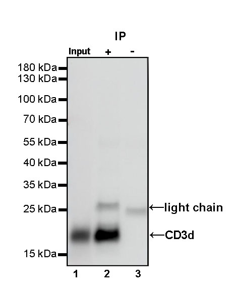
CD3d Rabbit mAb at 1/50 dilution (1 µg) immunoprecipitating CD3d in 0.4 mg Jurkat whole cell lysate.
Western blot was performed on the immunoprecipitate using CD3d Rabbit mAb at 1/1000 dilution.
Secondary antibody (HRP) for IP was used at 1/400 dilution.
Lane 1: Jurkat whole cell lysate 20 µg (Input)
Lane 2: CD3d Rabbit mAb IP in Jurkat whole cell lysate
Lane 3: Rabbit monoclonal IgG IP in Jurkat whole cell lysate
Predicted MW: 19 kDa
Observed MW: 21 kDa
免疫组化
IHC shows positive staining in paraffin-embedded human tonsil. Anti-CD3d antibody was used at 1/250 dilution, followed by a HRP Polymer for Mouse & Rabbit IgG (ready to use). Counterstained with hematoxylin. Heat mediated antigen retrieval with Tris/EDTA buffer pH9.0 was performed before commencing with IHC staining protocol.
免疫细胞化学
ICC shows positive staining in Jurkat cells. Anti-CD3d antibody was used at 1/500 dilution (Green) and incubated overnight at 4°C. Goat polyclonal Antibody to Rabbit IgG - H&L (Alexa Fluor® 488) was used as secondary antibody at 1/1000 dilution. The cells were fixed with 100% ice-cold methanol and permeabilized with 0.1% PBS-Triton X-100. Nuclei were counterstained with DAPI (Blue). Counterstain with tubulin (red).
Negative control: ICC shows negative staining in Raji cells. Anti-CD3d antibody was used at 1/500 dilution and incubated overnight at 4°C. Goat polyclonal Antibody to Rabbit IgG - H&L (Alexa Fluor® 488) was used as secondary antibody at 1/1000 dilution. The cells were fixed with 100% ice-cold methanol and permeabilized with 0.1% PBS-Triton X-100. Nuclei were counterstained with DAPI (Blue). Counterstain with tubulin (red).


评论(0)