


 大包装询价
大包装询价 产品介绍 引用文献(1) 评论(0)
宿主来源
Rabbit抗原名称
CD8α分子别名
Lyt-2免疫原
Synthetic Peptide细胞定位
Cell membraneAccession
P01731克隆号
SDT-142-52抗体类型
Rabbit mAb应用
IHC-P, FCM, WB, IF反应种属 ?
Ms纯化方式
Protein A浓度
0.5 mg/ml性状
Liquid缓冲体系
PBS, 40% Glycerol, 0.05%BSA, 0.03% Proclin 300
储存条件
12 months from date of receipt / reconstitution, -20 °C as supplied
| 应用 | 稀释度 |
|---|---|
| FCM | 1:50 |
| WB | 1:1000 |
| IF | 1:1000 |
| IHC-P | 1:500 |
CD8A encodes the CD8 alpha chain of the αβT cells, proposed as a quantifiable indicator for CD8+ CTL recruitment or activity assessments and a robust biomarker for responses to anti-PD-1/PD-L1 therapy.In NK-cells, the presence of CD8A homodimers at the cell surface provides a survival mechanism allowing conjugation and lysis of multiple target cells. CD8A homodimer molecules also promote the survival and differentiation of activated lymphocytes into memory CD8 T-cells.
免疫印迹
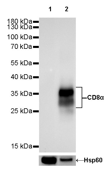
WB result of CD8α Rabbit mAb
Primary antibody: CD8α Rabbit mAb at 1/1000 dilution
Lane 1: RAW 264.7 whole cell lysate 20 µg
Lane 2: mouse thymus lysate 20 µg
Negative control: RAW 264.7 whole cell lysateSecondary antibody: Goat Anti-Rabbit IgG, (H+L), HRP conjugated at 1/10000 dilution
Predicted MW: 29 kDa
Observed MW: 28~37 kDa
Exposure time: 60s
流式分析
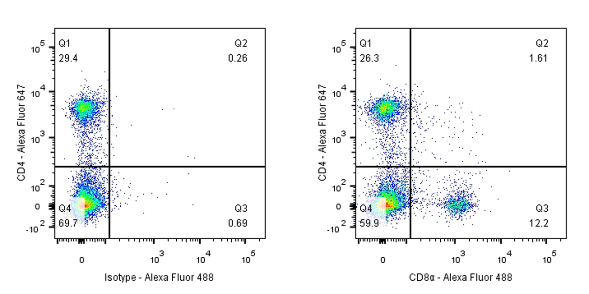
Flow cytometric analysis of mouse primary splenocytes labeling CD8α antibody at 1/50 (1 μg) dilution / (right panel) compared with a Rabbit IgG, Isotype Control / (left panel). Goat Anti-Rabbit IgG Alexa Fluor® 488 was used as the secondary antibody.
Cells were surface stained with CD4-Alexa Fluor® 647, then stained with rabbit IgG (Left) / CD8α (Right) separately. CD4 and CD8α are mutually exclusive expressed in mouse spleen. Gated on total viable cells.
免疫组化
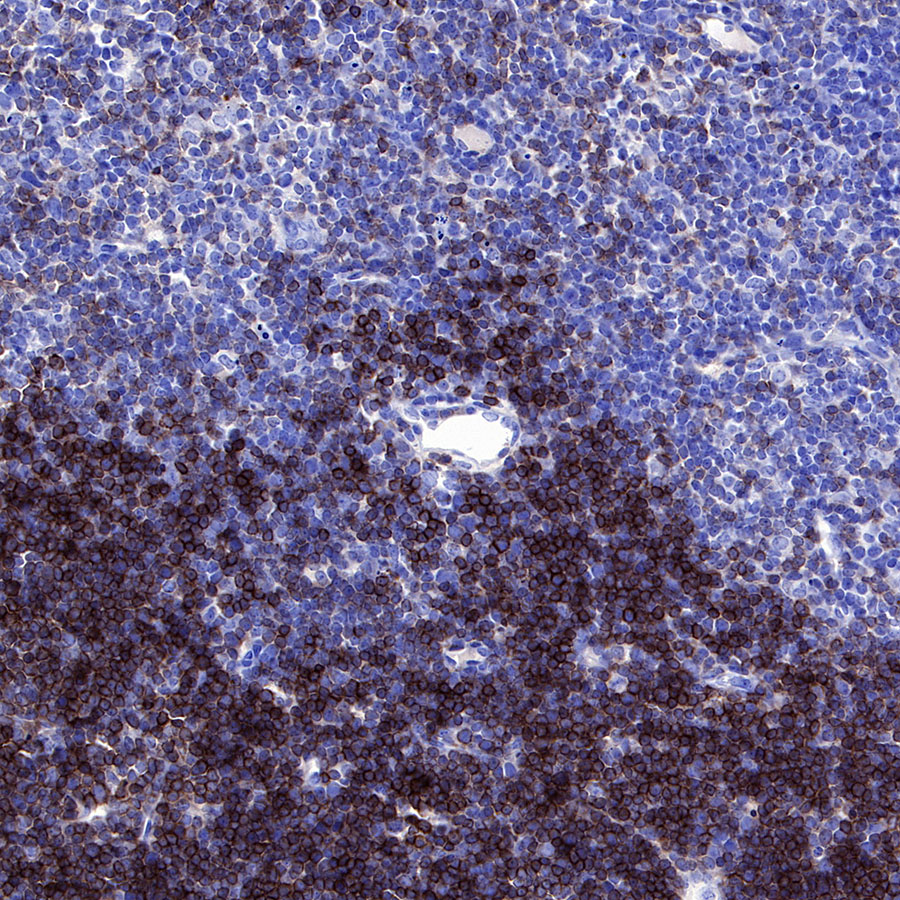
IHC shows positive staining in paraffin-embedded mouse thymus. Anti-CD8α antibody was used at 1/500 dilution, followed by a HRP Polymer for Mouse & Rabbit IgG (ready to use). Counterstained with hematoxylin. Heat mediated antigen retrieval with Tris/EDTA buffer pH9.0 was performed before commencing with IHC staining protocol.
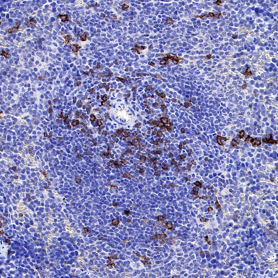
IHC shows positive staining in paraffin-embedded mouse spleen. Anti-CD8α antibody was used at 1/500 dilution, followed by a HRP Polymer for Mouse & Rabbit IgG (ready to use). Counterstained with hematoxylin. Heat mediated antigen retrieval with Tris/EDTA buffer pH9.0 was performed before commencing with IHC staining protocol.
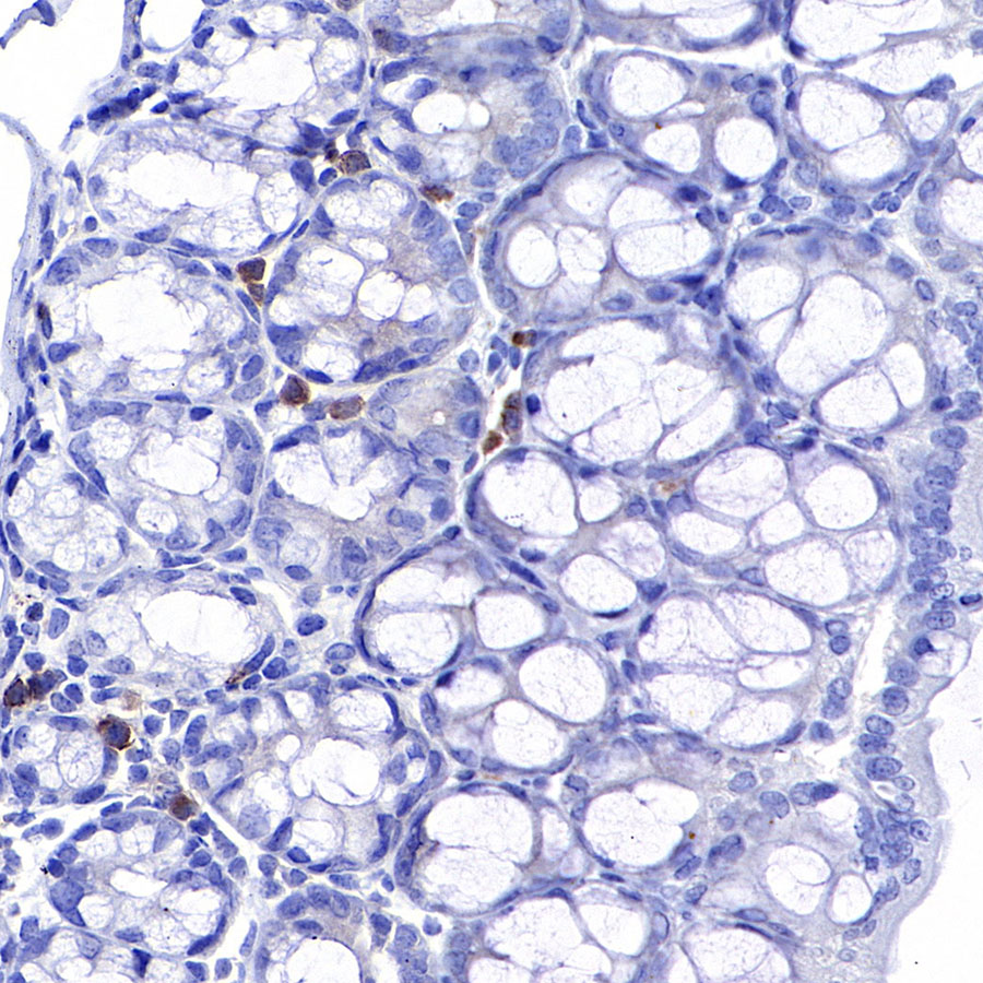
IHC shows positive staining in paraffin-embedded mouse colon. Anti-CD8α antibody was used at 1/500 dilution, followed by a HRP Polymer for Mouse & Rabbit IgG (ready to use). Counterstained with hematoxylin. Heat mediated antigen retrieval with Tris/EDTA buffer pH9.0 was performed before commencing with IHC staining protocol.
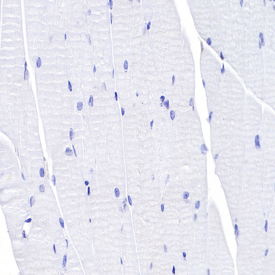
Negative control: IHC shows negative staining in paraffin-embedded mouse cardiac muscle. Anti-CD8α antibody was used at 1/500 dilution, followed by a HRP Polymer for Mouse & Rabbit IgG (ready to use). Counterstained with hematoxylin. Heat mediated antigen retrieval with Tris/EDTA buffer pH9.0 was performed before commencing with IHC staining protocol.
免疫荧光
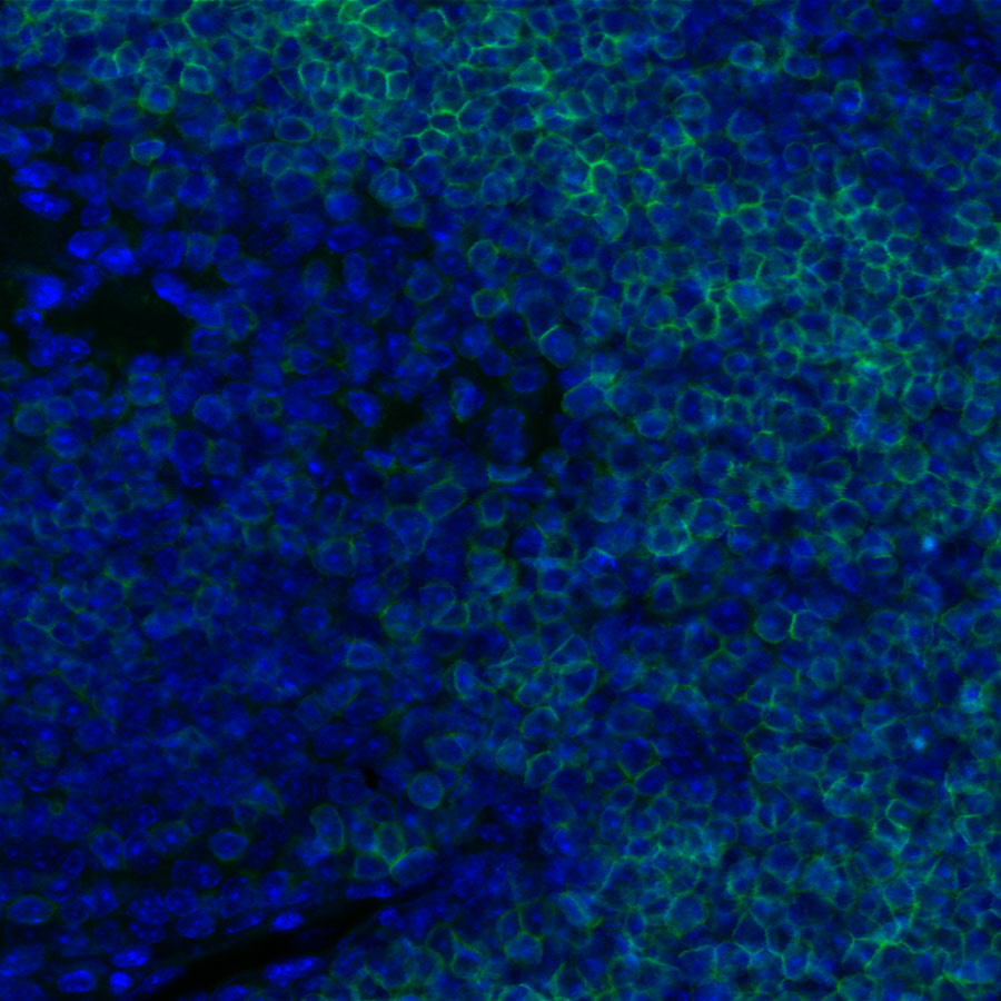
IF shows positive staining in paraffin-embedded mouse thymus. Anti-CD8α antibody was used at 1/1000 dilution and incubated overnight at 4°C. Goat polyclonal Antibody to Rabbit IgG - H&L (Alexa Fluor® 488) was used as secondary antibody at 1/1000 dilution.Heat mediated antigen retrieval with Tris/EDTA buffer pH9.0 was performed before commencing with ICC staining protocol. Nuclei were counterstained with DAPI.
引用文献(1)
- Fc–Fc interactions and immune inhibitory effects of IgG4: implications for anti-PD-1 immunotherapies
Zhang W, Chen X, Chen X, Li J, Wang H, Yan X, Zha H, Ma X, Zhao C, Su M, Hong L.
Journal for Immunotherapy of Cancer. 2024 Dec 01 ; 6
影响因子: 10.9
货号:S0B0034产品名称:CD8α Recombinant Rabbit mAb (SDT-142-52)


评论(0)